Can an MRI without contrast detect a brain tumor?

Daniel Johnson
Published Apr 04, 2026
Cranial computed tomography (CT) and magnetic resonance imaging (MRI) with and without contrast media are widely used for primary diagnosis of brain tumors. Standard T1- and T2-weighted MRIs detect brain tumors with high sensitivity.
Can an MRI without contrast detect a tumor?
MRI without contrast cannot generally help in evaluating the given tumor condition. MRI images with contrast are clearer than the images of MRI without contrast. Due to the high clarity of images gathered by MRI with contrast, they are easier for a medical specialist to evaluate and interpret.Can plain MRI detect brain tumor?
In general, diagnosing a brain tumor usually begins with magnetic resonance imaging (MRI). Once MRI shows that there is a tumor in the brain, the most common way to determine the type of brain tumor is to look at the results from a sample of tissue after a biopsy or surgery.What does an MRI without contrast of the brain show?
Non-contrast MRI is great option for patients for whom dye is not recommended, pregnant women and kidney-compromised patients. Non-contrast also provides greater images of blood vessel activity, detecting aneurysms and blocked blood vessels.Can a brain tumor be missed on an MRI?
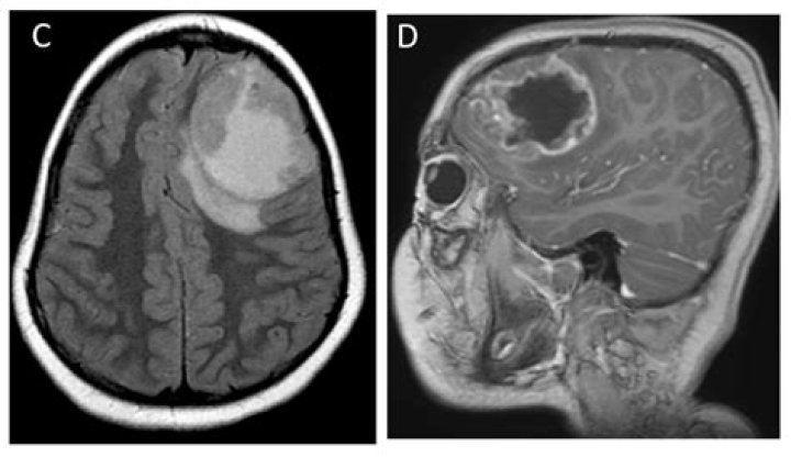
Imaging tests such as MRI and CT scans may show an abnormal area that is likely to be a brain or spinal cord tumor. But these scans can't always tell exactly what type of tumor it is. Often this can only be done by removing some of the tumor tissue in a procedure called a biopsy.Imaging brain tumors - 4 - Other low grade gliomas
Are MRI without contrast accurate?
Non-contrast MRI.A non-contrast MRI is also an effective exam for imaging your body's organs. Though it doesn't use contrast dye, it can still be quite accurate.
What were your first signs of a brain tumor?
Symptoms
- New onset or change in pattern of headaches.
- Headaches that gradually become more frequent and more severe.
- Unexplained nausea or vomiting.
- Vision problems, such as blurred vision, double vision or loss of peripheral vision.
- Gradual loss of sensation or movement in an arm or a leg.
- Difficulty with balance.
Can an MRI miss something?
A false negative diagnosis made off an MRI scan could lead the neurologist and patient down an incorrect path and delay an accurate diagnosis, or potentially miss it entirely. While MRI is not the only piece in the puzzle for MS diagnosis, it plays a significant role.Why would a neurologist order an MRI of the brain?
MRI is used to diagnose stroke, traumatic brain injury, brain and spinal cord tumors, inflammation, infection, vascular irregularities, brain damage associated with epilepsy, abnormally developed brain regions, and some neurodegenerative disorders.Which is better MRI or CT scan for brain?
MRI. CT scans are more widely used than MRIs and are typically less expensive. MRIs, however, are thought to be superior in regards to the detail of the image. The most notable difference is that CT scans use X-rays while MRIs do not.Can a brain tumor be detected without contrast?
Cranial computed tomography (CT) and magnetic resonance imaging (MRI) with and without contrast media are widely used for primary diagnosis of brain tumors. Standard T1- and T2-weighted MRIs detect brain tumors with high sensitivity.What can be mistaken for a brain tumor?
Brain tumour misdiagnosis can commonly be diagnosed as the following diseases, given the similarity across symptoms a patient suffers with: Alzheimer's disease. Encephalitis. Headaches or migraines.How do you rule out a brain tumor?
Diagnosis
- A neurological exam. A neurological exam may include, among other things, checking your vision, hearing, balance, coordination, strength and reflexes. ...
- Imaging tests. Magnetic resonance imaging (MRI) is commonly used to help diagnose brain tumors. ...
- Collecting and testing a sample of abnormal tissue (biopsy).
How accurate are MRI scans of the brain?
The overall diagnostic sensitivity and PPV were 72.0–90.7% and 91.9–95.4%. Diagnostic accuracy differed among tumor types. Some tumor types tended to be confused with each other. Preoperative MRI reports should not be relied upon too heavily in decision-making.How long does a brain MRI without contrast take?
The scan typically takes about 30 to 60 minutes. According to Cincinnati Children's Hospital and Medical Center, scans that don't require a contrast dye are generally shorter and may only take 30 to 45 minutes. Some procedures like the limited brain MRI only take about 5 minutes.What can an MRI tell a neurologist?
Some of the conditions a brain MRI can help diagnose or monitor include:
- A blood clot in your brain.
- Brain aneurysm.
- Brain hemorrhage.
- Brain infections (encephalitis).
- Brain damage associated with epilepsy.
- Brain tumors and cysts.
- Certain chronic neurological conditions, such as multiple sclerosis (MS).
- Dementia.
Can a neurosurgeon read an MRI?
Thirty-nine per cent of the psychiatrists and 11% of the neurologists only read the report but not the MRI images. Interestingly, no neurosurgeons reported reading only the report, but 7% declared they only read the MRI images. None of the other specialties, neurology or psychiatry, reported analysing only the images.Can MRI Miss brain aneurysm?
Aneurysm visible on MRI scan missedSadly there are cases in which medical practitioners have assessed the images of an MRI scan, yet have failed to identify an aneurysm, and thus failed to follow-up and treat the patient.